Friday, September 12, 2014

Light Independent Reaction

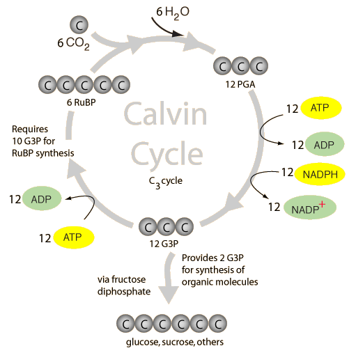
Image taken from: http://hyperphysics.phy-astr.gsu.edu/hbase/biology/imgbio/calvine.gif
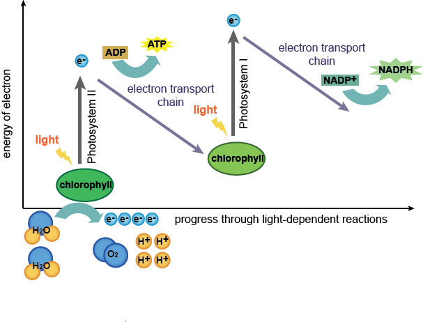
  • Happens in the stroma
  • Does not require light
  • ATP and NADPH from light dependent reaction is used to reduce carbon dioxide to triose phosphate and regenerate ribulose biphosphate (RuBP)
  • Carbon dioxide reacts with RuBP to form an unstable 6-carbon intermediate which breaks down into two molecules of 3 phosphoglycerate (PGA) catalysed by ribulose biphosphate carboxylase/oxygenase (Rubisco)
  • ATP and NADPH reduces PGA to form triose phosphate (G3P), forming ADP and NADP in the process
  • Most of the triose phosphate is used to regenerate RuBP
  • Some triose phosphate exit the cycle and is used to synthesise other products like starch and glucose
  • ADP and NADP are channeled back into the light dependent reaction to produce more ATP and NADPH


Wednesday, September 10, 2014

Light Dependent Reaction

Image taken from: http://www.shmoop.com/images/biology/biobook_photosyn_2.png
  • There are two photosystems at the thylakoid membranes of the chloroplast.
  • Photosynthetic pigments (eg. chlorophyll and carotenoids) are organized into the two photosystems, PSII and PSI.
  • Photosynthetic pigments absorb light energy which cause the excitation of their electrons (photoactivation).
  • Excited electrons are ejected from PSI and PSII due to their high energy.
  • The excited electrons produce adenosine triphosphate (ATP) from adenosine diphosphate (ADP) using ATP synthase.
  • Nicotinamide adenine dinucleotide phosphate (NADPH) is also produced fronicotinamide adenine dinucleotide phosphate ion (NADP+).
  • The electron transport chain and the ATP synthase complex at the thylakoid membrane are involved in the production of ATP and NADPH.
  • As light is involved in the production of ATP, it is known as photophosphorylation.
  • At PSII, the electrons used to produced ATP and NADPH are replenished by electrons from water molecules. 
  • Splitting of water (photolysis) results in the release of oxygen gas.
In simpler terms, when light falls on the thylakoid membrane,
  • Light causes photoactivation, which is the excitement of electrons by absorption of light energy, at the photosynthetic pigments at PSII and PSI.
  • Excited electrons are ejected from the reaction centers of PSII and PSI.
  • Ejected electrons are used to produce ATP and NADPH.
  • Electrons lost are replenished from photolysis of water.
  • Oxygen gas is released in the process.

Monday, September 8, 2014

Chloroplasts

Image taken from: https://benchprep.com/blog/wp-content/uploads/2012/08/chloroplast2.jpg

Image taken from: http://blog.canacad.ac.jp/bio/BiologyIBHL1/files/1477622.jpg


Chloroplasts
  • found in mesophyll cells
  • site for photosynthesis
  • surrounded by double membrane called chloroplast envelope
  • thylakoids are fluid-filled sacs
  • a stack of thylakoids is called a granum
  • stroma is a gel-like fluid that surrounds the grana
  • stroma contains soluble enzymes of the Calvin cycle, sugars, organic acids, etc.
  • excess carbohydrates from photosynthesis is stored as starch grains in the chloroplast
  • the two main photosynthetic pigments, chlorophyll and carotenoids, are embedded at the surface of thylakoid membranes
Chlorophyll
  • converts light energy to chemical energy
  • absorb mainly red and blue-violet light 
  • reflects green light which gives leaves their green colour
Carotenoids
  • yellow, orange, red or brown pigments
  • absorb strongly in blue-violet light

Sunday, September 7, 2014

Leaf Structure

Image taken from: https://encrypted-tbn1.gstatic.com/images?q=tbn:ANd9GcRMdbgJi7OECdEIZltQeEN-4XlpJUQnYaSeXWwa6ikEDcItbmgP


Cuticle 
  • thin waxy layer of lipids
  • prevents water loss through evaporation
  • allows light to pass through
Epidermis
  • transparent layer of cells
  • allows light to pass through
  • protects the cells underneath from damages
Palisade Mesophyll Cells
  • contains a large number of chloroplasts
  • tightly packed together vertically to allow as many cells as possible to absorb light
Spongy Mesophyll Cells
  • contains few chloroplasts
  • loosely packed with intercellular air spaces for fast diffusion of carbon dioxide
Stoma
  • surrounded by two guard cells
  • allows exchange of gases 
Vascular Bundle (Vein of leaf)
  • made up of xylem and phloem vessels
  • xylem transports water and mineral salts
  • phloem transports nutrients

Friday, September 5, 2014

Inhibitors

Many drugs and poisons are inhibitors of enzymes in the nervous system.

Competitive Inhibitor
  • resembles shape of substrate closely
  • competes with substrate for active site
  • binds to active site either reversibly or irreversibly
  • prevents substrate from binding to enzyme active site, reducing rate of reaction
Non-competitive Inhibitor
  • has a different structure from substrate
  • binds to an allosteric site, causing a change in the shape of active site
  • substrates can no longer bind to the active site and no enzyme-substrate complex can be formed

Factors affecting enzyme reactions

Substrate concentration
  • At low substrate concentration, increasing the substrate concentration will result in a proportional increase in the rate of reaction.
  • As there are sufficient active sites, the increase in substrate concentration will increase the frequency of effective collisions between the enzyme and substrate molecules, increasing the rate of formation of enzyme-substrate complex.
  • However, at high substrate concentration, increasing the substrate concentration will no longer speed up the reaction.
  • All the active sites of the enzyme molecules will be occupied at any given moment.
  • Any added substrates have to ‘wait’ until existing enzyme-substrate complexes are dissociated and the active sites become available for binding.
  • The rate of reaction can only be increased with the addition of enzymes.

Image taken from: http://alevelnotes.com/content_images/i73_Image3.gif

Enzyme concentration
  • At low enzyme concentration, increasing the enzyme concentration will result in a proportional and linear increase in the rate of reaction.
  • The increase in enzyme concentration provides more active sites which the substrate molecules can bind to, increasing the rate of effective collision and the formation of enzyme substrate complex.
  • However, at high enzyme concentration, increasing the enzyme concentration will no longer have effect on the reaction as all the substrate molecules would already have been converted into their products.
  • The rate of reaction can only be increased with the addition of substrates.
Image taken from: http://alevelnotes.com/content_images/i74_Image4.gif

Temperature
  • Enzymes work best at their optimum temperature, which is usually 40°C.
  • Below the optimum temperature, the rate of reaction increases linearly with the increase in temperature.
  • As the temperature increases, kinetic energy of the molecules increase. This increases the rate of effective collisions between enzyme and substrate molecules and the formation of enzyme-substrate complex.
  • Beyond the optimum temperature, the rate of reaction decreases even though the frequency of collisions increases.
  • Thermal agitation of the enzymes break the hydrogen bonds, ionic bonds and hydrophobic interactions that stabilize the specific 3D conformation of the enzyme.
  • The enzyme becomes denatured as the shape of the enzyme's active site is altered and is no longer complementary to that of the substrate.
Image taken from: http://alevelnotes.com/content_images/i71_gcsechem_18part2.gif

pH
  • Enzymes work best at their optimum pH level.
  • A change in pH level will result in the alteration of the ionic charge of the R groups of the amino acid residues.
  • This breaks the ionic bonds and hydrogen bonds that are responsible for maintaining the specific 3D conformation of the enzyme.
  • The enzyme becomes denatured as the shape of the enzyme's active site is altered and is no longer complementary to that of the substrate.
Image taken from: http://alevelnotes.com/content_images/i72_enzyme_ph_graph.gif


Sunday, June 22, 2014

Enzymes

What are enzymes?

  • large biological molecules which catalyse chemical reactions in living organisms
  • responsible for the metabolic processes which sustain life
  • enzymes can be proteins or ribonucleic acid (RNA)
Properties of enzymes:
  • lower the minimum amount of energy needed to start a reaction (activation energy)
  • has active site of specific shape, which is complementary to its substrate 
  • does not alter properties of end products of reaction
  • highly efficient in small amounts
  • denatured easily due to high heat
How do enzymes work?
  1. After an effective collision between a substrate and an enzyme (where the substrate binds to the active site of the enzyme), an enzyme-substrate complex is formed. 
  2. The substrate molecule is held in the active site by interactions such as hydrogen and ionic bonds between the R groups of the amino acids and the substrate molecule. 
  3. Enzyme catalyses conversion of substrate to the end product. 
  4. The alteration of chemical conformation results in the product being released from the active site since it is no longer complementary to the structure of the active site. 
  5. The active site is then available for other substrates to bind to it.
"Lock and Key" Hypothesis
  • there is an exact fit between the substrate and the active site of the enzyme
  • the enzyme (lock) has a unique shape complementary to the substrate (key)
  • enzymes are very specific; only substrates that are exactly complementary to its active site are able to bind with it
image taken from: http://katysstudynotes.files.wordpress.com/2010/11/enzymes.png

Induced Fit model
  • active site of enzyme is flexible 
  • active site is able to mould itself around the substrate to make the fit better
  • active site returns to original shape after the products are released 
Illustration of the induced fit model
image taken from: http://zebrafish.umdnj.edu/Pre-Enrollment/Resources/Biochemistry/Enzymes%20and%20Metabolism/Images/enzyme_fit_diagram.png


Food Tests

Benedict's Test for Reducing Sugars
  • Pour 2cmof solution sample solution into a test tube and add an equal volume of Benedict's solution. 
  • Shake the mixture.
  • Heat test tube in a boiling water bath for 5 minutes.
  • Record observation after 5 minutes.
Colour of precipitate and the corresponding amount of reducing sugar present
http://brilliantbiologystudent.weebly.com/uploads/1/4/8/3/14836282/44628.jpg?513

Biuret's Test for Proteins
  • Add 2cmof sample solution to a clean test tube and add 2cmof dilute sodium hydroxide solution.
  • Shake the mixture.
  • Add 1% copper (II) sulphate solution, drop by drop. Shake mixture after every drop and observe the colour change. 
  • If mixture remains blue, no protein is present.
  • If mixture turns violet/purple, protein is present. 
Iodine Test for Starch
  • Add 2cmof sample solution to a clean test-tube, followed by a few drops of iodine solution.
  • Observe the colour change.
  • If mixture remains brown, no starch is present.
  • If mixture turns blue black, starch is present.
Ethanol Emulsion Test for Fats
  • Add 2cmof sample solution to a clean test tube and add 2cmof ethanol.
  • Shake mixture thoroughly.
  • Add 2cmof water into the test tube and shake. 
  • Record the observation.
  • If mixture remains clear, no lipid is present.
  • If a white emulsion was obtained, lipid is present. 

Condensation and Hydrolysis

Condensation and hydrolysis are reverse processes of each other.

Condensation (or dehydration synthesis) is a chemical process which by 2 molecules are combined to make a larger molecule through the loss of water. It is the process which by biomolecules are formed.

Hydrolysis is a chemical process which by a macromolecule is broken down into smaller molecules by the addition of water. Often, enzymes are needed to carry out hydrolysis, as water alone is insufficient.

image taken from: http://classconnection.s3.amazonaws.com/311/flashcards/1406311/jpg/polymers1334535675092.jpg


Friday, June 20, 2014

Biomolecules — Lipids

What are lipids?
  • lipids are insoluble in water
  • contain C, H and O
  • contain less oxygen than carbohydrates
  • made up of fatty acids and glycerol
What are fatty acids?
  • contain hydrophobic "tail" of a long hydrocarbon chain, and hydrophilic "head" which is a carboxyl group
  • can be saturated (no double bonds, no kinks) or unsaturated (double bonds and kinks in fatty acid tail)
Classification of lipids
  • simple lipids
  • compound lipids
  • steroids and sterols
Simple Lipids
  • formed by joining fatty acid with an alcohol (eg. glycerol)
  • fats (eg. triglyceride) are simple lipids
  • triglyceride structure: glycerol + 3 fatty acid tails
Compound Lipids (eg. phospholipids)
  • found in phospholipid bilayer of cell membrane
  • phospholipid structure: phosphate head + 2 fatty acid tails

Biomolecules — Carbohydrates

Carbohydrates are the most abundant biomolecule in nature. 


Function of carbohydrates
  • building blocks for larger molecules 
  • energy storage — starch (plant) and glycogen (animal)
  • structural — cellulose cell wall (plant) and chitin (insects, crabs, shrimps)
The bond between monomers of carbohydrates is known as glycosidic bond.

Monosaccharides
  • monomers of carbohydrates
  • eg. glucose, fructose, galactose
  • all monosaccharides are reducing sugars
Disaccharides
  • made up of two monosaccharides

http://i.imgur.com/2Kx8x.jpg
Polysaccharides
  • made up of many monosaccharides
What is starch? 
  • polymer of glucose molecules
  • has two components; amylose and amylopectin
  • both fit together to form a complex 3-dimensional structure which is insoluble in water
  • amylose helices are entangled in the branches of amylopectin molecules
  • each amylose chain is coiled into a helix, with six glucose residues for every complete turn of the helix — compact shape making it a complex structure for storage
  • amylopectin have many branches

What is glycogen?
  • animal equivalent of starch
  • found in liver and skeletal muscles of vertebrate animals 

Starch and glycogen (energy stores)
  • their molecules have many side branches where glucose molecules can be removed from their tips (by enzymes)
  • their insolubility stops them interfering with osmosis
  • their compactness provides an efficient way to store lots of glucose for future cellular respiration

Cellulose (long, unbranched chain)
  • most abundant organic molecule on Earth
  • major component of cell wall in plants
  • made from long, straight unbranched chains of glucose
  • chains cross-linked by H-bonds which holds them tightly together (excludes water)
  • chemically very inert and insoluble 
  • many molecules form strong fibrils
  • only some bacteria, fungi and a very small number of animals can secrete cellulase enzymes

Tuesday, March 4, 2014

Biomolecules — Proteins

Amino Acids:

The basic building blocks of proteins are amino acids. Amino acids are the monomers which form polypeptide and proteins.

Properties of Proteins:
  • sensitive to pH and heat
  • shape determines function
  • enzymes are special group of proteins
  • contain H, N, C and S
  • made up of amino acids
There are about 20 essential amino acids found in proteins, and they differ in their R group. 

Amino acids join together to form a polypeptide through the process called condensation.

Two amino acids are joined by a peptide bond to form a dipeptide via condensation. Water is formed as a by product. 

The reaction is between an amino group and a carboxyl group. 

Continued condensation leads to the addition of more amino acids and thus resulting in a polypeptide chain.


Structure of proteins:
  1. Primary structure --> specific linear order which amino acids join to form a polypeptide chain
  2. Secondary structure --> regular folding of segments by hydrogen bonds (α-helix and β-pleated sheet)
  3. Tertiary structure --> (only for globular proteins) overall shape of polypeptide resulting from interactions* between the R group 
  4. Quaternary structure --> association of 2 or more polypeptide chains 
*Bonds that form tertiary structures:
  • hydrogen bonds
  • ionic bonds
  • disulfide bonds
  • hydrophobic interaction
Classification of proteins (based on structure):
  • Globular --> compact and spherical structure which is soluble
  • Fibrous --> long polypeptide chains twisted around each other, forming long fibres that provide tensile strength, insoluble 

Sunday, February 16, 2014

Cell Transport

Passive transport (requires no ATP as molecules move along a concentration gradient):
  • Simple Diffusion
  • Facilitated Diffusion
  • Osmosis
Active transport (requires ATP as molecules move against a concentration gradient):
  •  Endocytosis
  •  Exocytosis
  •  Pinocytosis (“drinking”)
  •  Phagocytosis
Simple Diffusion
  • Net movement of molecules in and out of the cell along a concentration gradient
  • Eg. gas exchange during photosynthesis, gas exchange at the alveoli 
Facilitated Diffusion
  • Movement of specific molecules down a concentration gradient through specific carrier proteins
  • Molecules that move by facilitated diffusion include glucose and amino acids
Osmosis

  • Net movement of water molecules from a region of higher water potential to a region of lower water potential, across a partially permeable membrane
  • Eg. absorption of water by plant roots


credits to: http://www.biologymad.com/resources/diffusionrevision.pdf

Cell membrane structure

Function of cell membranes:
  •      To compartmentalize the cell --> separate different enzymes of different organelles and prevent autolysis
  •      Control entry and exit of substances
  •      Increases surface area for exchange of substances
  •      Cell recognition
  •      Cell communication
  •      Site of chemical reactions (eg. light reactions of photosynthesis)


Fluid mosaic model


The cell membrane consists of:
  • phospholipids --> head is polar while tail is non-polar 
  • proteins --> peripheral/integral proteins 
  • glycoproteins (consists of carbohydrate chains bound to peripheral proteins and hydrophilic regions of integral proteins) 
  • cholesterol --> maintains membrane fluidity


Cell Structure — Animal cell vs Plant cell

Animal Cell structure


Plant Cell structure

credits to: http://www.diffen.com/difference/Animal_Cell_vs_Plant_Cell


Biological Molecules - You Are What You Eat: Crash Course Biology #3


A helpful video on biomolecules!


 ← screengrab from the video!